
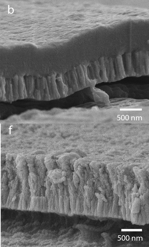
The top scanning electron microscope image (b) shows a cross section of the bioactive hydroxyapatite/YSZ coating without heat treatment. Note how the two layers are distinct. The bottom image (f) shows the coating after heat treatment. Note how the layers are now integrated. Image credit: North Carolina State University
(Visited 30 times, 1 visits today)