
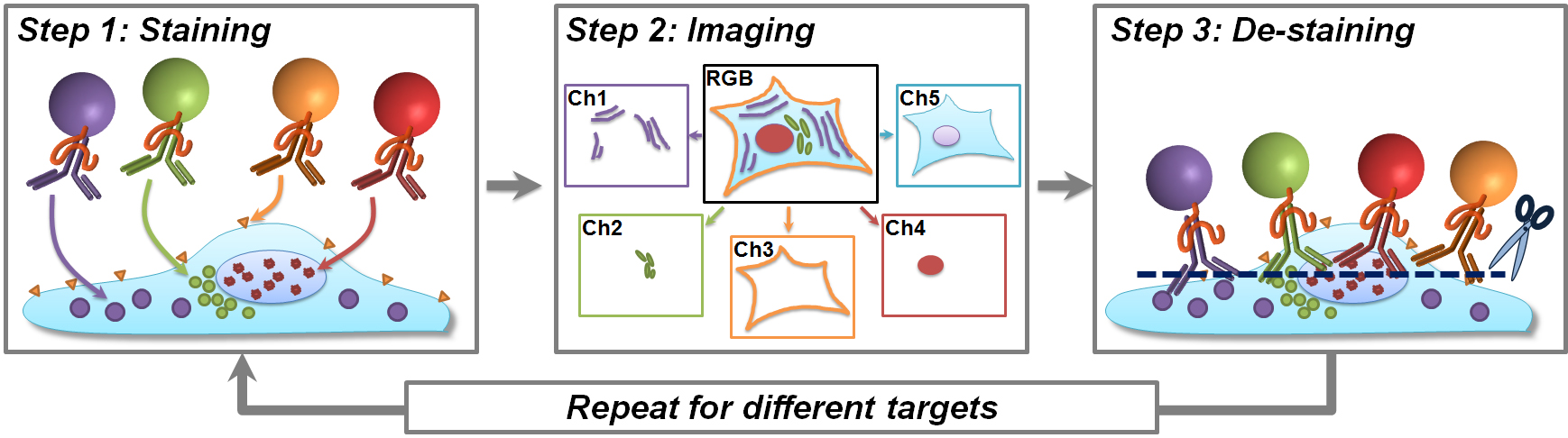
This figure shows the cyclical process developed in the study. In step 1, the colored balls representing quantum dots of different colors are used to label biomarkers in cell and tissue samples. Step 2 shows how each biomarker can be isolated and separated into distinct images for analysis. Step 3 illustrates how the tissue sample is flushed clean between rounds to begin biomarker testing again. Image credit: Xiaohu Gao (Click image to enlarge)
(Visited 26 times, 1 visits today)