
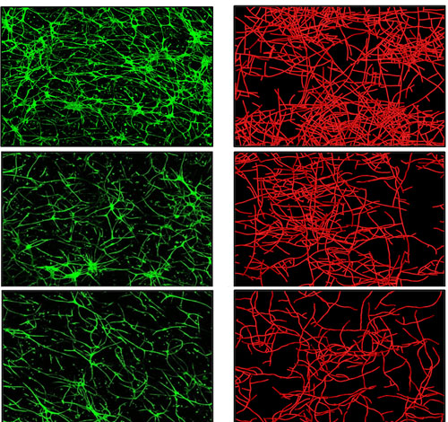
The microscope images on the left (green) show real blood vessels growing in culture, while the images on the right (red) are from a computer simulation of blood vessel growth. The top images show real and simulated blood vessel growth when vessel fragments are placed in an “extracellular matrix” of collagen with a relatively low density. The middle and bottom images show how blood vessel growth is impeded when they are placed in collagen matrix with medium and higher density, respectively. University of Utah bioengineers say the computer simulation of blood vessel growth is an early step toward new treatments to provide better blood supply to skin grafts and implanted ligament and tendon, as well as tissues damaged by diabetes and heart attack. Photo Credit: Jeff Weiss and Lowell Edgar, University of Utah